The signs and symptoms of NMOSD can be mistaken for MS1

~4 out of 10 patients with NMOSD are initially
misdiagnosed with MS1

Icon for optic neuritis Icon for optic neuritis Icon for optic neuritis

MRI scan showing a dense Gd-enhancing lesion at the posterior part of the right optic nerve in optic neuritis, a differential factor in diagnosing NMOSD vs MSMRI scan showing a dense Gd-enhancing lesion at the posterior part of the right optic nerve in optic neuritis, a differential factor in diagnosing NMOSD vs MS

NMOSD MRI5
Dense Gd-enhancing lesion at
the posterior part of the right
optic nerve

MRI scan showing an extensive Gd-enhancing lesion at the bilateral posterior part of the optic nerve/chiasm in bilateral optic neuritis, a clinical presentation observed in NMOSD vs MSMRI scan showing an extensive Gd-enhancing lesion at the bilateral posterior part of the optic nerve/chiasm in bilateral optic neuritis, a clinical presentation observed in NMOSD vs MS

NMOSD MRI5
Extensive Gd-enhancing lesion at the
bilateral posterior part of the optic
nerve/chiasm

Clinical Presentation MS NMOSD
Vision loss Check icon Check icon
Blindness Check icon
MRI Unilateral optic neuritis Unilateral or bilateral optic neuritis
Transverse Myelitis Transverse Myelitis Transverse Myelitis
Clinical Presentation MS NMOSD
Spasms Check icon Check icon
Limb weakness Check icon Check icon
Sensation loss Check icon Check icon
Paralysis Check icon Check icon
Loss of bladder/ bowel function Check icon Check icon
MRI Longitudinal, short-length spinal cord lesion LETM involving >50% of the spinal cord, centrally

MRI scan showing longitudinally extensive transverse myelitis extending from the brain stem at the area postrema to T2, a clinical presentation observed in NMOSD vs MS

Sagittal T2 image of the cervical spinal cord shows LETM extending from the brain stem at the area postrema to T2

Brain and brain stem Brain and brain stem Brain and brain stem

Sagittal T2-weighted area postrema MRI in an NMOSD patientSagittal T2-weighted area postrema MRI in an NMOSD patient

NMOSD MRI
Sagittal T2-weighted MRI of the area postrema

Clinical Presentation MS NMOSD
Hiccups Check icon
Nausea Check icon
Vomiting Check icon
MRI Ovoid, white matter lesions Periventricular and circumventricular lesions

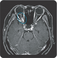
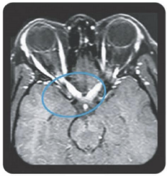
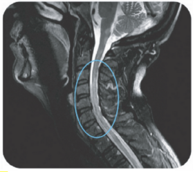
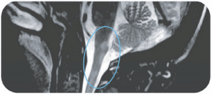
Area postrema syndrome is a core clinical characteristic of NMOSD that can appear both at disease onset and throughout the clinical course7

  • The spectrum of NMOSD symptoms is broad and includes nausea, vomiting, and intractable hiccups7,8
    • 4 out of 10 NMOSD attacks of the area postrema include all 3 symptoms7
  • Due to symptom severity, ~80% of patients with NMOSD-related area postrema syndrome are hospitalized7

Gd, gadolinium; LETM, longitudinally-extensive transverse myelitis.

  1. Beekman J, Keisler A, Pedraza O, et al. Neuromyelitis optica spectrum disorder: patient experience and quality of life. Neurol Neuroimmunol Neuroinflamm. 2019;6:1-14. doi:10.1212/NXI.0000000000000580
  2. National Multiple Sclerosis Society Website. Accessed November 21, 2022. https://www.nationalmssociety.org/Symptoms-Diagnosis
  3. NORD rare disease database. Accessed on November 21, 2022. https://rarediseases.org/rare-diseases/neuromyelitis-optica/
  4. Dutra BG, da Rocha AJ, Nunes RH, Martins Maia Júnior AC. Neuromyelitis optica spectrum disorders: spectrum of MR imaging findings and their differential diagnosis. RadioGraphics. 2018;38:169-193.
  5. Kim HJ, Paul F, Lana-Peixoto MA, et al. MRI characteristics of neuromyelitis optica spectrum disorder: an international update. Neurology. 2015;84:1165-1173. doi:10.1001/jamaneurol.2015.1276
  6. de Carvalho Jennings Pereira WL, Reiche EMV, Kallaur AP, Kaimen-Maciel DR. Epidemiological, clinical, and immunological characteristics of neuromyelitis optica: a review. J Neurol Sci. 2015;355:7-17.
  7. Shosha E, Dubey D, Palace J, et al. Area postrema syndrome: frequency, criteria, and severity in AQP4-IgG–positive NMOSD. Neurology. 2018;19:e1642-e1651.
  8. Sellner J, Boggild M, Clanet M, et al. EFNS guidelines on diagnosis and management of neuromyelitis optica. Eur Neurol. 2010;17:1019-1032.